
PDF chapter test TRY NOW
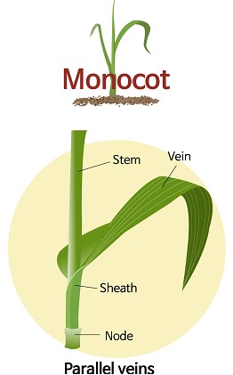
Monocot leaf
When you see externally, parallel veins are observed on the monocot leaf, which is different from the dicot leaf. There are unique features for its internal structure also, which is explained in this topic.
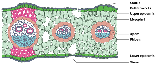
Transverse section of monocot leaf
The above picture is showing a sector of the transverse section of a monocot leaf or isobilateral leaf.
The following structures are seen in the section.
i. Epidermis:
The outermost epidermal layer is present on the upper side of the leaf and the lower side of the leaf. It is a single layer of parenchymatous cells without intercellular spaces. The outer walls of the cells are covered with the protective layer, the cuticle. Stomata is present on both upper and lower epidermis layers. Some parenchymatous cells on the upper epidermis are large and thin-walled. They are known as bulliform cells. Bulliform cells make the leaves curl during water stress and helps to reduce water loss due to evaporation.ii. Mesophyll:
Between the upper and lower epidermis, there is an entire mass of ground tissue called mesophyll. Unlike dicot leaf, no differentiation is observed in the mesophyll as palisade and spongy parenchyma. The cells are made up of parenchyma and are irregularly arranged with intercellular spaces. These cells contain chloroplasts and can take part in the process of photosynthesis.iii. Vascular bundles:
A large number of vascular bundles are present. Some of the vascular bundles are small, and some are large. Each vascular bundle consists of xylem and phloem and is surrounded by a sheath of cells made up of parenchyma called bundle sheath. The vascular bundle is conjoint, collateral and closed where the xylem is positioned towards the upper epidermis layer and the phloem is positioned towards the lower epidermis layer.Video explaining the internal structure of monocot leaf in comparison to dicot leaf
 Register for free to see more content