
PUMPA - SMART LEARNING
எங்கள் ஆசிரியர்களுடன் 1-ஆன்-1 ஆலோசனை நேரத்தைப் பெறுங்கள். டாப்பர் ஆவதற்கு நாங்கள் பயிற்சி அளிப்போம்
Book Free Demo“I think; therefore, I am.”
This is a famous statement made by the French philosopher Descartes. The process of thinking made him realise that he existed. Thinking and reasoning are essential things that give all the advances for humankind. It is done by a special type of tissue called nervous tissue or neural tissue. It also functions to stimulate muscle contraction, create awareness of the environment, and create emotions and memory.
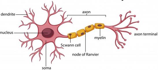
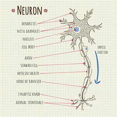
A neuron is a tiny structure found in the brain, spinal cord, and nerves. A single neuron can be up to a metre long and is made up of three parts:
- Cell body
- Axon
- Dendrites
Neuron anatomy
1. Cell body
The cell body is found in the grey matter of the brain’s central nervous system, but a few ganglia are located outside the CNS. It is also called cyton or soma.
Neurons have a star-shaped location on their cell bodies, with different hair-like elements extending from the edge. The cell body carries genetic information and helps maintain the neuron’s structure, also providing energy to drive activity.
Like other cell bodies, a neuron’s soma comprises a nucleus, cytoplasm, and specialised organelles such as mitochondria, golgi bodies, endoplasmic reticulum, ribosomes, and lysosomes. In neurons, the centrosome is absent. It is covered by a membrane that also permits it to interact with its surrounding environment.
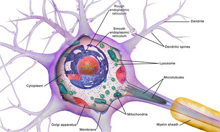
Cell body of neuron
The granular bodies found in the cell bodies are known as Nissl's granules.
Important!
Note: The Nissl's granules are irregular aggregates of rough endoplasmic reticulum containing a large number of attached and free ribosomes and polysomes. These granules are most likely responsible for the cell’s protein production.
Neuron
2. Axon
A single long, cylindrical conducting fibre extending from a neuron is known as an axon.
Axon terminals are fragile hair-like structures. Each branch ends in a synaptic knob, which is a bulb-like structure. Synaptic vesicles, which contain substances known as neurotransmitters, are found in the synaptic knob. Axons carry nerve impulses from the cell body to a synapse or neuromuscular junction.
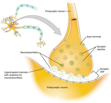
Chemical Synapse in axon terminal
3. Dendrites
These are short branched, hair-like fibres of a neuron. Nissl's bodies, neurofibrils and mitochondria are present in dendrites.
Most neurons have multiple dendrites, which extend outward from the cell body and are specialised to receive chemical signals from the axon terminal of other neurons. These signals are converted into tiny electric impulses by dendrites, transporting them inside toward the cell body.
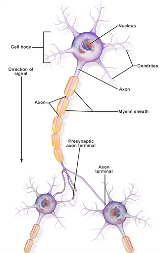
The image depicts the communication between a neuron's axon terminals and the dendrites of another neuron.
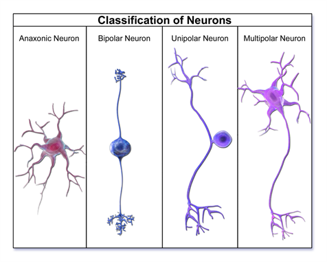
Types of neurons
There are four groups of neurons based on their functional existence to the structure.
- Apolar neurons
- Unipolar neurons
- Bipolar neurons
- Multipolar neuron
Classification of neurons
1. Apolar neuron:
They don't have any polarity. Axons and dendrites are not distinguished in these fibres. All fibres are of the same kind and can carry information towards or away from the cell body.
E.g. Neurons of a hydra.
E.g. Neurons of a hydra.
2. Unipolar neuron:
They have one axon and one dendrite only. The flow of information is unidirectional. These are sensory neurons. They can be found in invertebrate and vertebrate embryos.
3. Bipolar neuron:
Bipolar neurons are with one dendron and one axon at opposite poles. These neurons have a unidirectional information flow. They can be found in the retina of the eyes, as well as the olfactory epithelium.
4. Multipolar neuron:
One axon extends from the cell body and then split into two in a pseudo unipolar neuron. E.g. Dorsal root of ganglion.
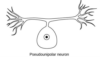
Pseudo unipolar neuron
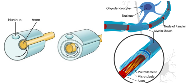
Types of axons:
Axon forms nerve fibres, and they are of two types.- Myelinated nerve fibres
- Non-myelinated nerve fibres
1. Myelinated nerve fibres:
Schwann cells envelop myelinated nerve fibres, forming the myelin sheath around the axon. The nodes of Ranvier are the spaces between two neighbouringmyelin sheaths. These fibres are present in the spinal and cranial nerves.
2. Non-myelinated nerve fibres:
A Schwann cell that does not form a myelin sheath around the axon surrounds a non-myelinated nerve fibre. There are no Ranvier nodes. These fibres can be found in both the autonomic and somatic nervous systems.
Non-myelinated and Myelinated nerve fibres
Reference:
https://commons.wikimedia.org/wiki/File:Neuron_Cell_Body.png
https://www.freepik.com/premium-vector/diagram-neuron-anatomy_2480497.htm#page=1&query=neuron&position=33
https://commons.wikimedia.org/wiki/File:1225_Chemical_Synapse.jpg
https://commons.wikimedia.org/wiki/File:Neuron_Part_1.png
https://commons.wikimedia.org/wiki/File:Neuron_Classification.png
https://commons.wikimedia.org/wiki/File:Figure_35_01_04.jpg
https://en.wikipedia.org/wiki/Central_nervous_system#/media/File:Periferal_nerve_myelination.jpg
https://en.wikipedia.org/wiki/Myelin#/media/File:Neuron_with_oligodendrocyte_and_myelin_sheath.svg
 Register for free to see more content