
PUMPA - SMART LEARNING
எங்கள் ஆசிரியர்களுடன் 1-ஆன்-1 ஆலோசனை நேரத்தைப் பெறுங்கள். டாப்பர் ஆவதற்கு நாங்கள் பயிற்சி அளிப்போம்
Book Free DemoIn a scary situation, our heart beats quicker, the breathing rate will increase, thus supplying our muscles with more oxygen, reducing the blood flow to the digestive system and skin.
All of these responses work together to prepare the animal's body to deal with the event. But this coordination of events is controlled by the hormone called adrenaline which is secreted by the adrenaline glands.
Adrenal glands
The paired structures on the top of the kidneys are known as the adrenal glands. These glands are also known as emergency glands. An outer cortex and an inner medulla make up each adrenal gland.

Adrenal gland
(a) Adrenal cortex
The embryonic mesoderm gives rise to the adrenal cortex. There are three layers to the adrenal cortex.
- Zona glomerulosa
- Zona fasciculata
- Zona reticularis
1. Zona glomerulosa: It's the area just below the capsule's outer zone. Mineralocorticoids are a hormone secreted by this gland.
2. Zona fasciculata: It is located in the central zone and consists of cells that release glucocorticoids and are arranged in long straight columns.
3. Zona reticularis: It is the inner zone, and it is made up of cells that emit sex corticoids and are grouped in branching cords.
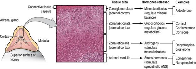
Composition of the adrenal gland and the hormones they release
(b) Adrenal Medulla
The adrenal medulla develops from the embryo's neuroectoderm. Chromaffin cells make up the medulla of the adrenal gland. Adrenaline (epinephrine) and noradrenaline (norepinephrine) are two hormones secreted by the adrenal medulla. Catecholamines are the general name for these hormones.
Epinephrine and norepinephrine are derived from the amino acid called tyrosine. These hormones are termed emergency raising of hairs, sweating, and other things because they are secreted quickly in reaction to stress and during an emergency.
The hormones adrenaline and noradrenaline both raise the heart rate, the strength of cardiac contractions, and the speed of respiration. They also aid in the breakdown of glycogen into glucose, which raises blood glucose levels. These hormones also aid the breakdown of fats and proteins.
Important!
The adrenal glands are sometimes known as "glands of emergency" because of the hormones that release.
The video explaining the location and structure of adrenal glands
Some cortical hormones are mentioned below.
Mineralocorticoids
They maintain the sodium and potassium ion ratio, that regulates electrolytic and fluid balance.
One of the most important mineralocorticoids in humans is aldosterone (salt-retaining hormone). Aldosterone helps the kidneys with active sodium reabsorption, passive water reabsorption, and potassium excretion.
Glucocorticoids
- These hormones are released in response to stress and when our body's equilibrium of nutrition is disturbed.
- Glucocorticoids not only affect the carbohydrate metabolism, but they also affect protein and fat metabolism.
- Gluconeogenesis is aided by glucocorticoids. The process of gluconeogenesis is the production of glucose from a non-carbohydrate source. As a result, they maintain glucose homeostasis.
- They also slow down protein synthesis and promote the formation of fat droplets from lipids. Immunity is suppressed by glucocorticoids, which reduce the number of eosinophils and lymphocytes that produce antibodies.
- Cortisol, corticosterone, and cortisone are the three major hormones found in glucocorticoids.
Cortisol is one of the most well-known glucocorticoids. Cortisol's main functions are listed below.
- It stimulates the liver to produce carbohydrates from non-carbohydrates such as amino acids and glycerol.
- Cortisol also causes a rise in the concentration of amino acids in the blood and increases the breakdown of proteins within cells.
- Cortisol's third action is to promote fat breakdown in adipose tissue and the release of fatty acids into the bloodstream. Cortisol is a stress hormone that our bodies produce when we are under stress. Hence, it is also known as the stress hormone.
Sexcorticoids or sex hormones:
Both male and female sex hormones are found in sexcorticoids.
- Androgens are male sex hormones that are vital in the development of a male foetus.
- Estrogens are female sex hormones produced by the adrenal cortex and are responsible for developing female secondary sexual characteristics.
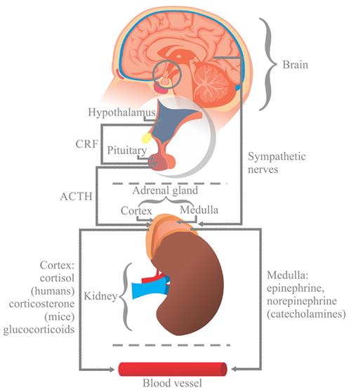
Pathway of release of hormones from adrenal cortex and medulla
Disorders related to the activity of the adrenal gland
The following are some of the most common adrenal cortical disorders:
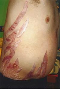
Addison’s disease
Hyposecretion of the adrenal cortical hormone causes Addison’s disease. Mineralocorticoid deficiency affects electrolytic balance, causes dehydration, and results in low blood sugar and bronze skin pigmentation.

The above picture shows the hyposecretion of adrenal cortical hormone - Addison's disease
Cushing’s syndrome
The person affected with Cushing's syndrome
Conn’s Syndrome (Aldosteronism)
Excess aldosterone increases the sodium-potassium ratio, which raises blood pressure. Excess water retention also raises blood pressure. Muscle weakness and cramping, as well as paralysis, are common symptoms.
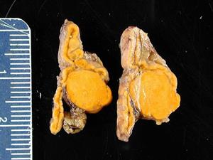
Adrenal gland affected by Conn's syndrome due to hyperaldosteronism
Adrenal Virilism
In females, excessive sex corticoid secretion causes male features such as a beard, moustache, and a change in voice. Adrenal virilism is the medical term for this illness. Sexual maturity occurs when these hormones are secreted in excess at a young age.
Gynaecomastia
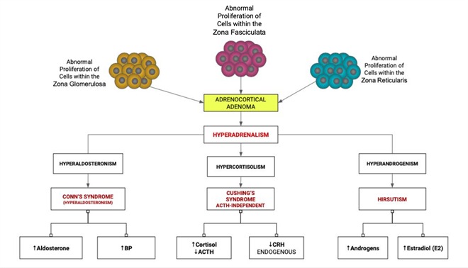
The flowchart of adrenocortical adenoma and its effects:
Pathology of adrenal cortex
Reference:
https://www.flickr.com/photos/143951580@N06/46556419855/in/photolist-2dW2PC4-9Ls4eL-doi3hJ-mpcFQW-5SCk1b-bNNtNT-5bQFni-4j7aVc-M6k7AD-8LSzA1-bXAMpq-6hdwiE-9P4MdW-9WbzHc-6sTBtJ-4aJYaD-8oY1Wd-CqGuoi-9wZAis-cfm2rd-eKqxGV-2bjXt7V-2i2NDgg-5ENC9J-eb9zpR-cFq6m-cFq6E-cFq6o-cFq6s-cFq6A-5EJjb4-5ENAvQ-5EJjUP-7KAqVW-bCKzNt-2iMT5c8-2iMZJ5V-5ENAcb-9uQi7r-5ENzSo-dVDaic-2T2vrw-4sYbpk-e9tkJB-ea2EaG-ea2E7J-e9z1vb-ea2E6C-e9tkNx-e9z1sw
https://upload.wikimedia.org/wikipedia/commons/c/ca/1818_The_Adrenal_Glands.jpg
https://commons.wikimedia.org/wiki/File:Response_to_stress.jpg
https://upload.wikimedia.org/wikipedia/commons/c/ca/1818_The_Adrenal_Glands.jpg
https://commons.wikimedia.org/wiki/File:Response_to_stress.jpg
https://upload.wikimedia.org/wikipedia/commons/thumb/9/93/Addison%27s_disease.jpg/512px-Addison%27s_disease.jpg
https://upload.wikimedia.org/wikipedia/commons/thumb/2/21/Straie_with_cushing_syndrome_new_photo.jpg/512px-Straie_with_cushing_syndrome_new_photo.jpg
https://upload.wikimedia.org/wikipedia/commons/5/50/Adrenal_Cortex_Pathology.png
https://upload.wikimedia.org/wikipedia/commons/thumb/0/04/Adrenal_gland_Conn_syndrome4.jpg/512px-Adrenal_gland_Conn_syndrome4.jpg
 Register for free to see more content