PDF chapter test TRY NOW
Small intestine
The longest part of the alimentary canal but has a small diameter. The length of the small intestine various from one organism to another based on the type of food it consumes. It is about \(6.25\)\(m\) in length.
Example:
Herbivores like a cow, deer that consume grass have a longer intestine as cellulose in grass takes longer to digest. On the other hand, meat is easier to digest, and thus carnivores like lion, tiger have shorter small intestine.
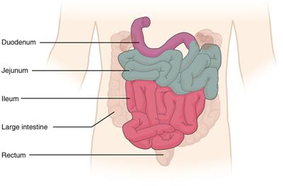
The small intestine is composed of three parts:
a. Duodenum - It is C-shaped and is as long as the breadth of \(12\) fingers. It contains foliate villi and mainly absorbs iron.
b. Jejunum - Middle part of the small intestine, which is thick-walled and more vascular.
c. Ileum - Longest part of the small intestine, which is thinner than the jejunum. It is about \(3.5m\) long. The ileum contains lymphatic nodules called Payer's patches that produce lymphocytes (WBC types).

Small intestine
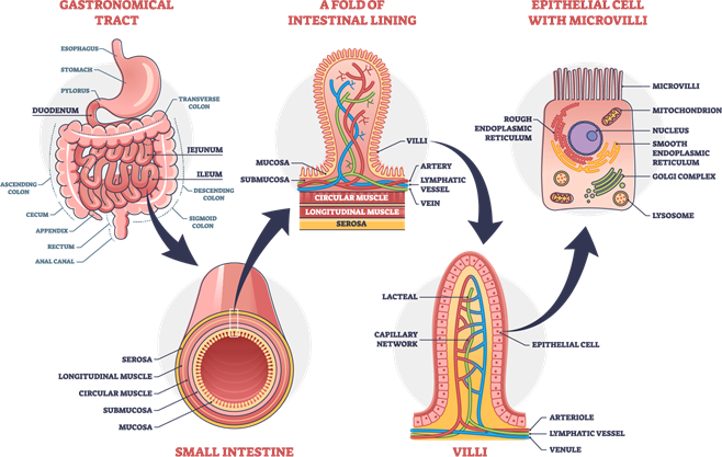
Both ileum and jejunum are highly coiled. Payer's patches are the distinguishing feature of the ileum. Villi (finger-like projections) are present in the small intestine except in the Payer's patches. Villi increase the surface area of the small intestine.
Each of the villus is covered with epithelium that contains lymph capillary and blood capillaries. The small intestine completely digests the proteins, carbohydrates, fats and nucleic acids. The absorption of nutrients into the blood and lymph occurs here.

Intestinal villi
The presence of blood capillaries in villi takes the absorbed nutrients to various parts of the body, which is utilised for obtaining ATP from glucose (glucose is oxidized to \(CO_2\), \(H_2O\) and ATP), the building of new tissues and repairing of old tissues. In addition, the small intestine secretes digestive enzymes, cholecystokinin, secretin, enterocrinin, enterogastrone.
Large intestine
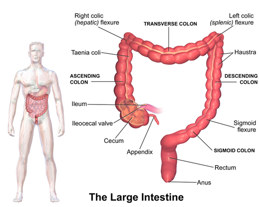
The large intestine diameter is larger than the small intestine. It is about \(1.5 metres\) in length and is divided into four parts:
a. Caecum and vermiform appendix - Caecum is a pouch-like structure whose walls contain prominent lymphoid tissue. The vermiform appendix is an outgrowth of the caecum. It is a vestigial part that is a slightly coiled blind tube.
b. Colon- Caecum leads to the colon and is divided into four regions:
(i) ascending colon
(ii) transverse colon
(iii) descending colon
(iv) sigmoid colon
Ascending colon is the part of the colon that is the shortest. The colon has small pouches called haustra.
c. Rectum- The sigmoid colon leads into the rectum. It comprises the last \(20 centimetres\) of the digestive tract and terminates in the long anal canal. The rectum acts as temporary storage of the undigested food.
d. Anus - Anus is the terminal part of the large intestine and the alimentary canal.
The undigested, semi-solid waste is expelled out from the body as faeces through the anus. This is called egestion.
Faeces are expelled from time to time from the anus out of the body. The anus has an internal anal sphincter composed of smooth muscle fibres and an external anal sphincter comprised of voluntary muscle fibres.
The function of the large intestine includes the absorption of water and the elimination of solid wastes. In addition, some amount of vitamin K and vitamin B complex are manufactured by bacteria in the large intestine.

Reference:
https://commons.wikimedia.org/wiki/File:2417_Small_IntestineN.jpg
https://commons.wikimedia.org/wiki/File:Blausen_0604_LargeIntestine2.png
