
PDF chapter test TRY NOW
Our human eye cannot able to see cells since they are much smaller. To see such smaller objects, the human eye requires the help of an instrument called a microscope. This topic addresses about the compound and an electron microscope.
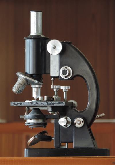
Compound microscope
It is an improved Hook’s microscope design, consisting of the eyepiece, body tube, objective lenses, coarse adjustment, fine adjustment, arm, stage, swivel, base, mirror, clip, microscope slide and condenser.
The word microscope can be split into two individual words, ‘micro’ and ‘scope’, where the term ‘micro’ refers to small and ‘scope’ refers to observe. This denotes that a microscope is an instrument used to observe small elements.
These are high-resolution instruments. They are used for observing very minute objects, e.g. cells, with the help of a microscope, the small size of a cell can be magnified up to \(300 - 1500\) times. Microscope used in schools is a compound microscope. It uses sunlight for illumination of objects to see. So it is called a light microscope. An electron microscope is used to watch the complex internal structure of the cell.
Compound microscopes have a combination of two lenses that amplify both magnifying powers as well as the resolving power. The specimen to be examined is generally mounted on a clear glass slide and place on the sample stage between the condenser lens and objective lens.
Application of Compound Microscope:
- A compound microscope is of prominent use in pathology labs to recognize infectious diseases.
- It assists in noticing and understanding the microbial world of bacteria and viruses, which is otherwise invisible to the naked eye.
- Plant cells are examined, and the microorganisms thriving on it can be ascertained with a compound microscope. Thereby, a compound microscope has proved to be crucial to biologists.
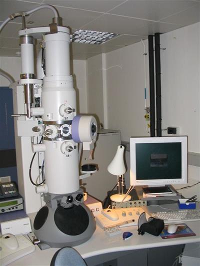
Electron microscope
Even though light microscopes are used, why do we need an electron microscope?
Objects smaller than the wavelength of light are difficult to be seen at higher resolution in a light microscope. However, electrons are used to image structures in an electron microscope, and electron beams have shorter wavelengths than light, allowing smaller objects to be seen at higher resolutions.
Electron microscope Applications of Electron microscope:
Electron microscope (EM) is used to produce high-resolution photographs of biological and non-biological specimens. It's used in biomedical research to look at the structure of tissues, cells, organelles, and macromolecular complexes in great detail.
Reference:
https://upload.wikimedia.org/wikipedia/commons/a/a5/Compound_Microscope_%28cropped%29.JPG
https://commons.wikimedia.org/wiki/File:Electron_Microscope.jpg
 Register for free to see more content