
PUMPA - SMART LEARNING
எங்கள் ஆசிரியர்களுடன் 1-ஆன்-1 ஆலோசனை நேரத்தைப் பெறுங்கள். டாப்பர் ஆவதற்கு நாங்கள் பயிற்சி அளிப்போம்
Book Free DemoStratified epithelial tissue has multiple layers of cells and is classified as
- Stratified columnar epithelial tissue
- Stratified squamous epithelial tissue
- Stratified cuboidal epithelial tissue
- Stratified ciliated columnar epithelial tissue
1. Stratified columnar epithelial tissue:
It is made up of tall, cylindrical pillar-like cells with multiple layers.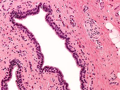
Stratified columnar epithelial tissue
Location: Linings of the urethra, a tube connects the kidneys and urinary bladder through which urine excretion happens.
Function: It helps to protect the underlying tissues. They often perform a role of limited secretion, depending on the tissues' location.
2. Stratified squamous epithelial tissue:
Location of these tissues are subjected to wear and tear. So, rapid regeneration of cell will occur.

Stratified squamous epithelial tissue
It is classified into two types.
a. Keratinised stratified squamous epithelium
b. Non-keratinised stratified squamous epithelium
a. Keratinised stratified squamous epithelium
Outer layers contains a protein called keratin, which is hard and waterproof
Location: Skin epidermis, hair, nails.

Keratinised stratified squamous epithelium
Function: This tissue gives protection from abrasion and infection. Also, it helps to prevent the drying of the underlying tissue.
b. Non-keratinised stratified squamous epithelium
Outer layers do not contain keratin. This tissue don't prevent water loss.
Location: Cornea, oral cavity, linings of oesophagus, rectum and internal portion of lips.

Non-keratinised stratified squamous epithelium
Function: Mucus is secreted by non-keratinized epithelia as an extra defensive and lubricating layer.
3. Stratified cuboidal epithelial tissue:
This tissue has cube-shaped cells with multiple layers.

Stratified cuboidal epithelial tissue
Location: Linings of sweat gland ducts, salivary and pancreatic ducts. The epidermis of fish is made of this tissue.
Function: They protect sweat gland ducts, mammary glands, and salivary glands.
4. Stratified ciliated columnar epithelial tissue:
This tissue has ciliated and cylindrical shaped cells present in the outer and basal layers of the tissue.

Stratified ciliated columnar epithelial tissue
Location: Linings of larynx and upper part of the soft palate.
Function: Secretion or absorption
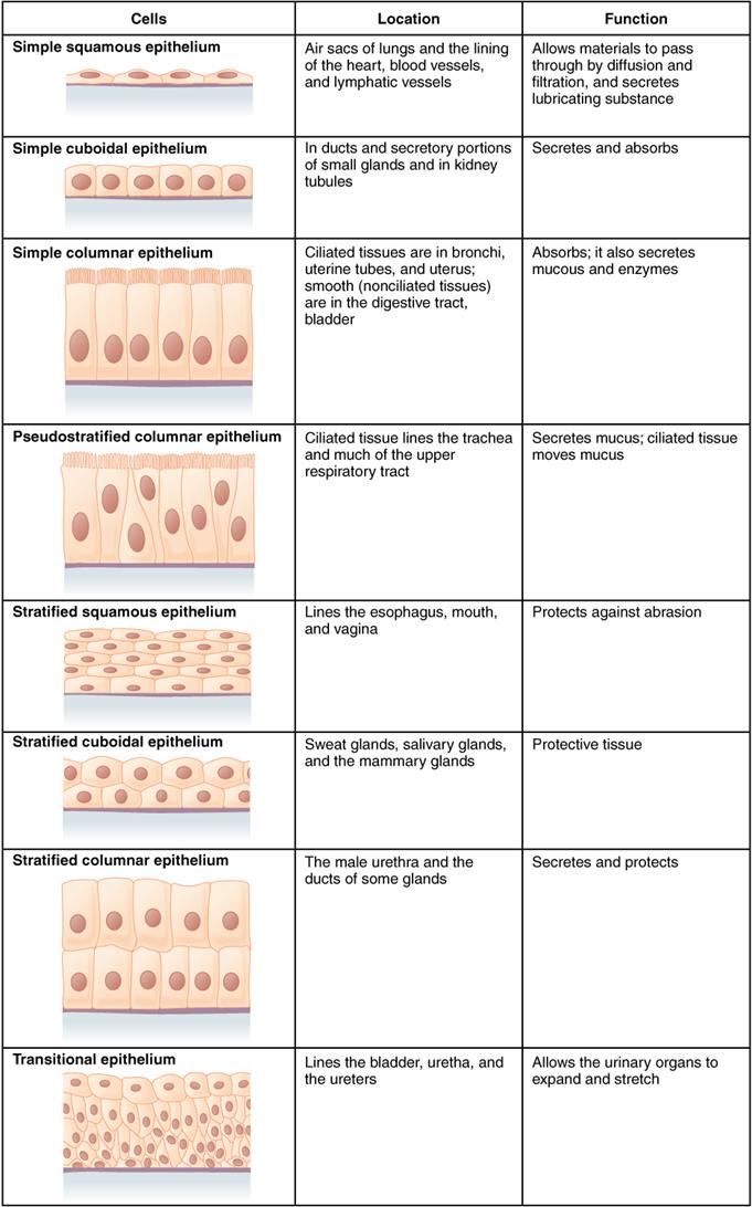
Summary of epithelial tissue cells
Glandular epithelial tissue:
Glands carry out the secretion of useful substances such as enzymes and hormones in an animal body. These are developed from epithelial tissue.
A portion of epithelial cells folds inward, which results in the formation of a multicellular gland. It is called glandular epithelium.
Location: It is found in the stomach, intestine and pancreas.
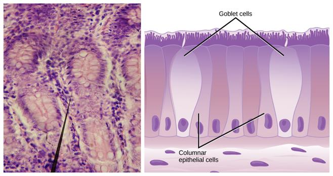
Glandular epithelial tissue
Function: It involves the synthesis and secretion of chemical substances at the epithelial surface.
Important!
Based on the epithelial layers
- Simple - Single layer
- Stratified - More than one layer
- Ciliated - Cells with cilia
Based on the cell shapes
- Squamous - Flat
- Cuboidal - Cube
- Columnar - Rectangular
Reference:
https://commons.wikimedia.org/wiki/File:Epithelial_Tissues_Stratified_Squamous_Epithelium_(40230838950).jpg
https://commons.wikimedia.org/wiki/File:Thuc_quan.jpg
https://upload.wikimedia.org/wikipedia/commons/c/c2/Stratified_cuboidal_epithelium_animated.gif
https://en.wikipedia.org/wiki/Epithelium#/media/File:423_Table_04_02_Summary_of_Epithelial_Tissue_CellsN.jpg
https://upload.wikimedia.org/wikipedia/commons/7/7e/Glandular_epithelium_of_small_intestine_%28crypts_of_Lieberk%C3%BChn%29.jpg
https://commons.wikimedia.org/wiki/File:Figure_33_02_03.jpg
 Register for free to see more content