
PUMPA - SMART LEARNING
எங்கள் ஆசிரியர்களுடன் 1-ஆன்-1 ஆலோசனை நேரத்தைப் பெறுங்கள். டாப்பர் ஆவதற்கு நாங்கள் பயிற்சி அளிப்போம்
Book Free DemoIn the above topic, we learnt about meristematic tissues. But plants can't do their functions with only meristematic tissues.
1. Plants have to store food and water
2. Plants have to get mechanical support
1. Plants have to store food and water
2. Plants have to get mechanical support
To perform such kind of specific functions, plants require permanent tissues.
Permanent tissue
A permanent tissue is made up of mature, living or dead cells divided from meristematic tissues.
Process of differentiation:
\(\text{Meristematic tissue}\) \(\xrightarrow{\small{\text{Differentiation process}}}\) \(\text{Permanent Tissues}\)
In plants, meristematic tissue loses its ability to divide. After they take up specific role.
This process of taking up a permanent shape, size, and a function of a meristematic tissue into a permanent tissue is called differentiation.
This process of taking up a permanent shape, size, and a function of a meristematic tissue into a permanent tissue is called differentiation.
Important!
Differentiation process causes the development of various types of permanent tissues.
There are two types of permanent tissue.
1. Simple Permanent tissue
2. Complex Permanent tissue
Simple Permanent Tissue
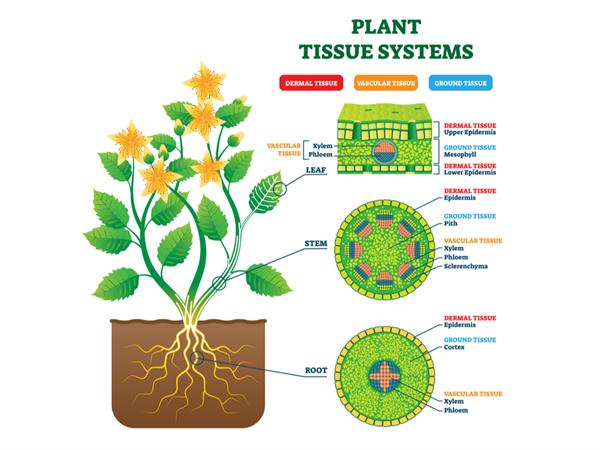
Plant tissue systems vector illustration. Anatomical diagram with leaf, stem and root microscopic graphic. Plant inner vascular, dermal and ground cross section
Simple permanent tissue consists of the same cells performing a similar function and occur in homogenous groups. They are not such specialized cells and with thin cell walls made up of cellulose. Cells are loosely spaced in this tissue. So intercellular spaces between these cells are large.
Location: Generally found in few layers below epidermis
Types of simple permanent tissues:
1. Parenchyma
2. Collenchyma
3. Sclerenchyma
2. Collenchyma
3. Sclerenchyma
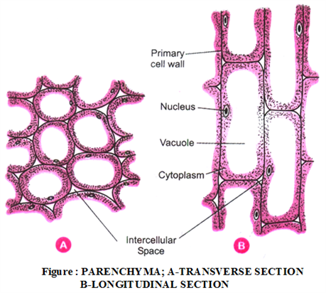
Parenchyma tissue
Parenchyma word derived from Greek (Para means"beside", enchyma means "in-filling"). The term parenchyma was coined by Grew. It is the simplest, unspecialized, most primitive, most abundant permanent tissue that evolved first; therefore, it is treated as a fundamental tissue. Framework of tissues in cortex, Pith, mesophyll of the leaves and floral parts are provided by parenchyma cells.
The shape of the parenchyma cells ultimately depends on their function.
- They are spherical shape in ground parenchyma.
- Irregular in spongy parenchyma.
- Cylindrical and elongated in palisade parenchyma.
- Parenchyma cells are elongated and armed in form in aerenchyma cells.
- These cells are loosely arranged with intercellular spaces.
- The cell wall is thin, which is made up of cellulose.
- It is a living cell with active protoplasts.
Functions of parenchyma:
1. It is responsible for storage of food, nutrients and water.
2. It provides mechanical support to the plants.
2. It provides mechanical support to the plants.
3. As it is a living tissue, metabolic activities occur in it.
4. Secondary tissues, responsible for the secondary growth of a plant is formed by the de-differentiation process of parenchyma.

Leaf tissue structure
Chlorenchyma: In some cases, parenchyma tissue contains chlorophyll and involves in the process of photosynthesis. This type of Parenchyma tissue is called as chlorenchyma.
Aerenchyma: In aquatic plants, parenchyma tissues have large air cavities inside it. These tissues float inside the water because of buoyancy caused by the air cavities. This type of parenchyma tissue is called as aerenchyma.
Example:
Water hyacinth and Elodea.

Aquatic plants of Water hyacinth and Elodea, Monocot stem cortex in Elodea
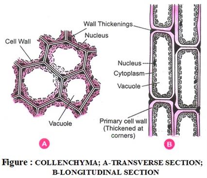
Collenchyma tissue
Collenchyma word derived from Greek (Kolla means glue). The term collenchyma was coined by Schleiden \((1839)\).
Location: Leaf Stalks below the epidermis and leaf midrib. It is absent in dicot roots, monocot plants and underground parts.
- The cells of these tissues are living cells.
- It has a central vacuole and peripheral cytoplasm with nucleus elongated and unevenly thickened at the corners because of the deposition of pectin and cellulose.
- Intercellular spaces between cells in this tissue is tiny.
Functions of collenchyma:
1. It gives flexibility and elasticity to the plant and prevents tearing of leaves.
2. It provides mechanical support for the plant.
3. This tissue enables parts of the plants such as tendrils and stems of climbers to bend easily without any breaking.
2. It provides mechanical support for the plant.
3. This tissue enables parts of the plants such as tendrils and stems of climbers to bend easily without any breaking.
Sclerenchyma tissue
Sclerenchyma word derived from Greek (Scleros means hard).
Location: In stems, around vascular bundles, in the veins of leaves, in the hard covering of seeds and nuts.
- The cells of this tissue are dead cells and act as a skeleton in plants.
- Cells are long and narrow in appearance with no nucleus and no cytoplasm.
- It is the most hardened plant tissue.
- Cell walls of this tissue are uniformly thickened due to lignin, which often provides no intercellular spaces between cells. E.g., Jute and coir fibres. They are rich in lignin.
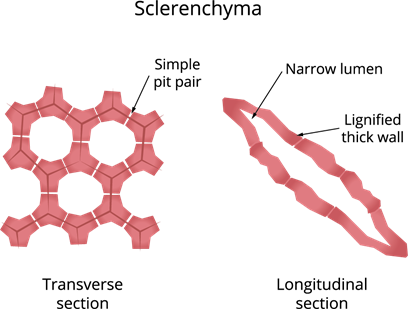
Sclerenchyma cells are of two types viz., Sclereids and Fibres .
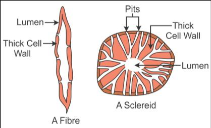
Sclereids: These are also called grit cells or stone cells. These are small cells, where the lumen is so tiny due to higher thickening of the cell wall as in drupe fruit (mango, coconut, walnut).
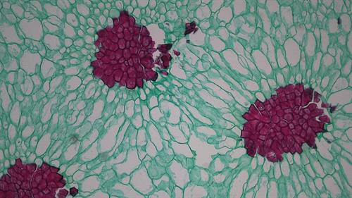
Sclereid cells in pear (Pyrus) fruit under the microscope
Fibres: They are very long, narrow, thick, lignified cells. Lumen is large as compared to the sclereids. In the thick wall, both the sclereids and fibres are present in thin areas called as pits. Pits are narrow and unbranched. These fibres are present in bundles and provide strength and hardness.
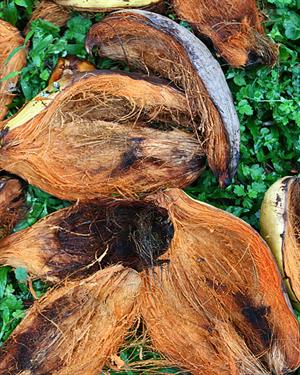
Functions of sclerenchyma: This tissue gives very rigid mechanical support to plants so that plants are hard and stiff. For example, husk of coconut is made up of sclerenchymatous tissue.
Importance of fibres and sclereids: It is used to manufacture of ropes, mats and certain textile fibres.
Reference:
https://www.flickr.com/photos/144872528@N06/29379783355
https://www.flickr.com/photos/144872528@N06/29092177320/in/photostream/
https://upload.wikimedia.org/wikipedia/commons/e/e7/Aquatic_Monocot_Stem_Cortex_in_Elodea_%2836915455834%29.jpg
https://commons.wikimedia.org/wiki/File:Elodea_canadensis_kz03.jpg
https://commons.wikimedia.org/wiki/File:Leaf_Tissue_Structure.svg
https://commons.wikimedia.org/wiki/File:Sclereids_in_Pyrus_pear_(36452689110).jpg
https://commons.wikimedia.org/wiki/File:Water_hyacinth_aerenchyma.JPG
 Register for free to see more content