
PDF chapter test TRY NOW
“I think; therefore, I am.”
This is a famous statement made by the French philosopher Descartes. The process of thinking made him realise that he existed. Thinking and reasoning are essential things that give all the advances for humankind. It is done by a special type of tissue called nervous tissue or neural tissue. It also stimulates muscle contraction, create awareness of the environment, and create emotions and memory.
A neuron is a tiny structure found in the brain, spinal cord, and nerves. A single neuron can be up to a metre long and is made up of three parts:
- Cell body
- Axon
- Dendrites
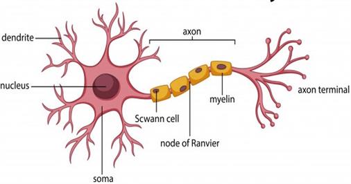
Neuron anatomy
1. Cell body:
The cell body is found in the grey matter of the brain’s central nervous system, but a few ganglia are located outside the CNS. It is also called cyton or soma or perikaryon.
Neurons have a star-shaped location on their cell bodies, with different hair-like elements extending from the edge. The cell body carries genetic information and helps maintain the neuron’s structure, also providing energy to drive activity.
A neuron’s soma or cyton comprises a central nucleus with abundant cytoplasm. This cytoplasm is called neuroplasm.
The cytoplasm consists of large granular bodies called Nissl's granules and some other specialised organelles such as mitochondria, golgi bodies, endoplasmic reticulum, ribosomes, and lysosomes. Inside the cytoplasm,numerous neurofibrils help to transmit the impulses to and from the cell body.
The cytoplasm is covered by a membrane that also permits it to interact with its surrounding environment. In neurons, the centrosome is absent, so they do not have the ability for cell division.

The cell body of a neuron
The granular bodies found in the cell bodies of a neuron are known as Nissl's granules.
Important!
Note:
The Nissl's granules are irregular aggregates of rough endoplasmic reticulum containing a large number of attached and free ribosomes and polysomes. These granules are most likely responsible for the cell’s protein production.

Neuron
2. Axon:

Schematic and microscopic view of nerve cells
A single long, cylindrical conducting fibre extending from a neuron is known as an axon.
- Axon terminals are fragile hair-like structures. Each branch ends with a bulb-like structure (swelling) called the synaptic knob. Synaptic vesicles, which contain substances known as neurotransmitters, are found in the synaptic knob.
- Axon's plasma membrane is called axolemma.
- Axon's cytoplasm is called axoplasm.
- Axon may or may not be covered by a protective sheath called the myelin sheath. It acts as an insulator and ensures rapid transmission of nerve impulses.
- Myelin sheath is covered by a layer of Schwann cells called neurilemma.
- Nodes of Ranvier are depressions in the myelin sheath that occurs at regular intervals and the region between the nodes is called an internode.
- Axons carry nerve impulses from the cell body to a synapse or neuromuscular junction.

Myelinatednerve cell and chemical synapse in the axon terminal
3. Dendrites:
These are short branched hair-like cytoplasmic processes of a neuron. Nissl's bodies, neurofibrils and mitochondria are present in dendrites.
Most neurons have multiple dendrites, which extend outward from the cell body and are specialised to receive chemical signals from the axon terminal of other neurons. These signals are converted into tiny electric impulses by dendrites, transporting them inside toward the cell body or cyton.

The image depicts the communication between a neuron's axon terminals and the dendrites of another neuron.
Reference:
https://commons.wikimedia.org/wiki/File:Neuron_Cell_Body.png
https://www.freepik.com/premium-vector/diagram-neuron-anatomy_2480497.htm#page=1&query=neuron&position=33
https://commons.wikimedia.org/wiki/File:1225_Chemical_Synapse.jpg
https://commons.wikimedia.org/wiki/File:Neuron_Part_1.png
https://commons.wikimedia.org/wiki/File:Figure_35_01_04.jpg
https://en.wikipedia.org/wiki/Central_nervous_system#/media/File:Periferal_nerve_myelination.jpg
https://en.wikipedia.org/wiki/Myelin#/media/File:Neuron_with_oligodendrocyte_and_myelin_sheath.svg
https://en.wikipedia.org/wiki/File:Nerve_cells.gif
 Register for free to see more content