
PDF chapter test TRY NOW
The thymus is partly an endocrine gland and partly a lymphoid organ. At birth, the thymus weighs between \(10\) and \(15grams\). It continues to grow till puberty, and after that, it diminishes in size until it weighs about \(10grams\).
It is located in the mediastinum (upper part of the chest) between the sternum and aorta.
It is a soft, bilobed and pinkish mass of lymphoid tissue.
The thymus secretes a hormone called thymosin that stimulates the development of certain types of WBCs involved in immunity.
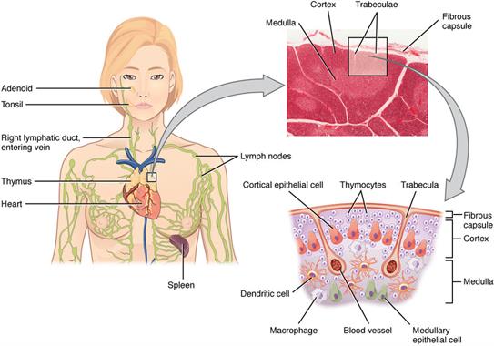
Histology and location of the thymus gland
Functions of thymosin:
- Thymosin has a stimulatory effect on immune function as it stimulates the development of WBCs.
- It stimulates the production and differentiation of lymphocytes.
- Thymosin also hastens the attainment of sexual maturity.
Pineal gland:
The pineal gland weighs around \(0.1 - 0.2\) grams. It is present between the cerebral hemispheres. It is a small rounded body that consists of pineal cells and supporting glial cells.
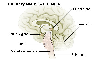
Location of pineal gland
The pineal gland contains the pinealocyte cells that secretes the hormone melatonin. The melatonin hormone is also called the "sleep hormone" as it promotes sleep. It is also called a "time messenger".
The secretion of melatonin differs according to the levels of light from the environment. The secretion of the hormone increases in dim light and decreases in bright light.
Melatonin concentration in the blood rises in the evening and at night, dropping low around the afternoon.
Thus, the pineal gland acts as a "biological clock" and signals nighttime information throughout the body. Exposure to light at night time decreases melatonin production, causing an interruption in sleep.
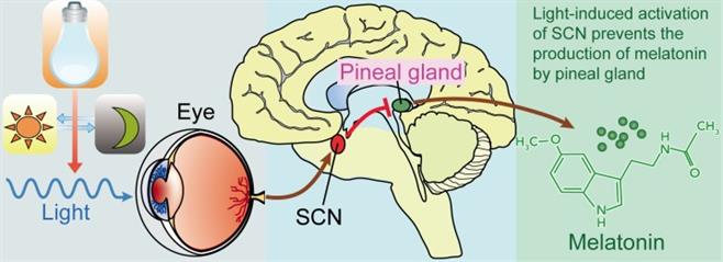
Activation of melatonin
Melatonin suppression has thus been linked to sleep disturbances and related metabolic disorders.
Reference:
https://upload.wikimedia.org/wikipedia/commons/thumb/9/99/2206_The_Location_Structure_and_Histology_of_the_Thymus.jpg/1024px-2206_The_Location_Structure_and_Histology_of_the_Thymus.jpg
https://upload.wikimedia.org/wikipedia/commons/4/43/Pituitary_pineal_glands.jpg
https://upload.wikimedia.org/wikipedia/commons/1/12/Light%2C_suprachiasmatic_nuclei_%28SCN%29%2C_and_the_pinealmelatonin_circuit.jpg
https://upload.wikimedia.org/wikipedia/commons/4/43/Pituitary_pineal_glands.jpg
https://upload.wikimedia.org/wikipedia/commons/1/12/Light%2C_suprachiasmatic_nuclei_%28SCN%29%2C_and_the_pinealmelatonin_circuit.jpg
 Register for free to see more content