
PUMPA - SMART LEARNING
எங்கள் ஆசிரியர்களுடன் 1-ஆன்-1 ஆலோசனை நேரத்தைப் பெறுங்கள். டாப்பர் ஆவதற்கு நாங்கள் பயிற்சி அளிப்போம்
Book Free DemoBefore studying the stages of sexual reproduction in humans, let us first look at the male and the female reproductive organs in humans. Human beings are unisexual and have two separate sexes - male and female. Both the sexes (male and female) are morphologically different from each other.
The reproductive system comprises of two different parts:
1. Primary reproductive sex organs - It includes the gamete producing gonads, i.e., the testes (in a male) and the ovaries (in female).
2. Secondary or accessory reproductive sex organs
Accessory sex organs:
The accessory sex organs include the glands, passages and other such associated structures. The accessory sex organs include:
- Vas deferens, epididymis, seminal vesicles, prostate gland, Cowper's gland (bulbourethral gland) and penis in the male.
- Fallopian tubes, uterus, cervix and vagina in the female.
The secondary sex organs are involved in:
- Process of ovulation
- Fertilization process where male and female gametes fuse
- Division of the fertilized egg until the formation of an embryo
- Pregnancy
- Development of the foetus
- Childbirth
Now let us focus on the primary reproductive organs, their cells and their role in reproduction in males and females.
Male Reproductive System:
The male reproductive system consists of :
1. A pair of testes that produces sperms.
2. A pair of sperm ducts called Vas deferens or Ductus deferens that carries the sperms. The vas deferens joins with the urethra that comes from the urinary bladder.
3. Accessory glands that contribute to the seminal fluid. The seminal vesicles secrete a fluid into the ejaculatory duct. The fluid, along with the sperms, is called semen. The prostate gland also pours its secretion to the urethra. This also mixes with the semen.
4. Penis that transfers sperms into the female.
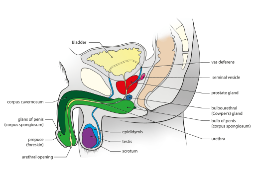
Male reproductive system
Testes:
Testes (singular testis) are the reproductive glands of the male that are present in a thin-walled sac called the scrotum. Each of the testes is an oval-shaped organ. It has a length of about \(4\ \)- \(5\) \(cm\) and a width of about \(2\) - \(3\) \(cm\). They lie outside the abdominal cavity of a man.
Now, we shall look into the cells that are present in a testis.
Each of the testes is covered by three layers:
1. Tunica vaginalis - the outer covering of the testis
2. Tunica albuginea - fibrous tissue that covers the testes
3. Tunica vasculosa - consists of a network of capillaries
Many septa from the tunica albuginea layer divide the testes into pyramidal lobules. Each testis has about \(250\) compartments of pyramidal lobules. In each lobule, the highly coiled seminiferous tubules, Sertoli cells, and the Leydig cells (interstitial cells) are present.
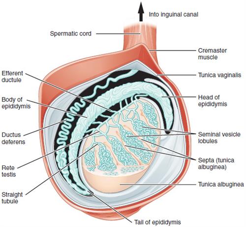
Longitudinal section of testes
The process of spermatogenesis (sperm formation) occurs in the seminiferous tubules. The Sertoli cells are the supporting or nursing cells that provide nutrients to the developing sperms. The cells also secrete another protein called the inhibin that suppresses the FSH synthesis.
The Leydig cells have a polyhedral shape and lie between the seminiferous tubules. The Leydig cells secrete the hormone testosterone. Testosterone is the hormone that initiates the process of spermatogenesis.
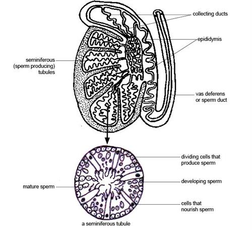
Anatomy and physiology of a seminiferous tubule
Reference:
https://commons.wikimedia.org/wiki/File:Male_genital_system_-_Sagittal_view.svg
https://commons.wikimedia.org/wiki/File:Figure_28_01_03.JPG
https://upload.wikimedia.org/wikipedia/commons/8/82/Anatomy_and_physiology_of_animals_The_testis_%26_a_magnified_seminferous_tubule.jpg
https://www.youtube.com/watch?v=k60M1h-DKVY&t=113s
 Register for free to see more content