
PUMPA - SMART LEARNING
எங்கள் ஆசிரியர்களுடன் 1-ஆன்-1 ஆலோசனை நேரத்தைப் பெறுங்கள். டாப்பர் ஆவதற்கு நாங்கள் பயிற்சி அளிப்போம்
Book Free Demo1. Oviducts/ Fallopian tubes:
These are a pair of muscular tubes of about \( 10 - 12\ cm\) in length. They connect each of the ovaries with the uterus. At the end of the ovary, the tube is broad and have a funnel-like structure called an infundibulum. The end of the tube is made up of several folds called fimbriae.
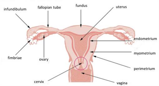
Female reproductive tract displaying infundibulum and fimbriae
The inner wall of the oviduct is ciliated that helps in the propagation of the egg. The fallopian tubes are the sites where fertilisation of the egg takes place.
2. Uterus:
It is a hollow, pear-shaped cavity broader on the upper end and narrower on the lower end. The uterus is present in the pelvic region behind the urinary bladder. The upper part is called the body or fundus of the uterus, and the lower end is called the cervix.
The cervix opens into the vaginal canal that opens to the outside. Cervix is made of a sphincter muscle that controls the opening and closing of the uterus.
The uterine wall has three layers. They include:
1. Innermost layer is the endometrium, made up of several glands and blood vessels. This layer undergoes morphological changes during menstruation or the menstrual cycle and the development of an embryo. The inner surface of the uterus is a site for the implantation of the embryo.
2. The middle layer is the myometrium, a smooth muscular layer composed of the major volume of the uterus wall.
3. The outer layer is the perimetrium.
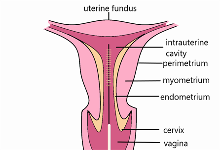
Uterine layers
3. Vagina:
The vagina is a \(9 \ cm\) long muscular tube lined with epithelial cells. The vaginal opening in young females is partially covered with a thin mucous membrane called the hymen. This is often broken early in females during strenuous work and physical activities like exercise, cycling, horse riding, etc. It receives the sperms and acts as a birth canal during childbirth.
4. Vulva:
Vulva is the external female genitalia. The vaginal opening has two pairs of folds on either side. The outer fold is thicker with hair, sweat glands and sebaceous glands and is called labia majora. The inner folds are thinner and devoid of hair. It is called labia minora.
Below is a video that explains the parts of the female reproductive system and emphasizes the functions of each:
Accessory glands:
There are two types of female reproductive glands. They include:
1. Bartholin's or large vestibular glands are small, round structures located at either side of the vaginal opening.
2. Mammary glands are present in the breasts of the females. Mammary glands help in lactation, i.e., the production of milk after childbirth.
Reference:
https://commons.wikimedia.org/wiki/File:Illu_cervix.jpg
https://commons.wikimedia.org/wiki/File:Labeled_tao_brush.png
https://www.youtube.com/watch?v=-5SOvWaW_OY
 Register for free to see more content