
PDF chapter test TRY NOW
The heart is a hollow, muscular pumping organ that pumps out the blood into the blood vessels. It is made up of cardiac muscles and is the size of our fist.
Important!
The human heart is present between the lungs, slightly tilted toward the left and above the diaphragm in the thoracic cavity.
Covering of the heart:
The heart consists of three layers:
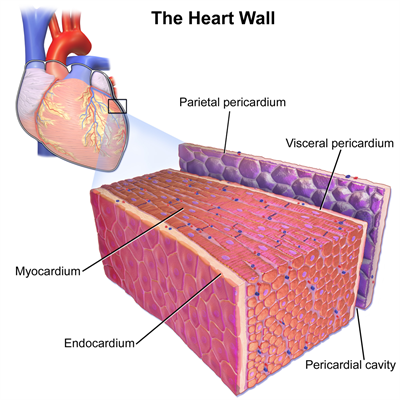
Internal structure of the heart wall
1. Pericardium - It is the outer double-walled sac. Between the layers, lubricating pericardial fluid is present. The pericardial fluid reduces the friction during the heartbeat and protects the heart from shocks and mechanical injuries.
2. Myocardium - Middle layer
3. Endothelium - Internal layer
The human heart has four different chambers that prevent the \(O_2\)-rich blood from mixing with the blood containing \(CO_2\). The four chambers include the:
- Right atrium
- Right ventricle
- Left atrium
- Left ventricle
Anatomy of the human heart
- The two upper thin-walled chambers are called auricle or atria (singular atrium).
- The two lower thick-walled chambers are called ventricles. Ventricles form the lower part of the heart.
What is a septum?
A wall or partition that separates the chambers of the heart is called a septum. It is made up of cardiac muscles. The septum prevents the mixing of oxygenated and deoxygenated blood.
The left and the right auricles are separated by an interatrial septum, while an interventricular septum separates the left and right ventricles. 
 1. Right atrioventricular valve:
Interatrial and interventricular septum
Important!
The left atrium is smaller than the right atrium.
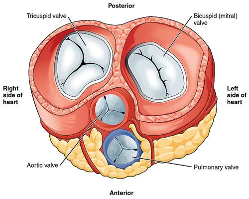
Valves:
Valves are muscular flaps that regulate the flow of blood in a single direction.
Why are the valves present in the heart?
1. To ensure a unidirectional flow of blood.
2. To prevent the flow of blood backwards during the contraction of atria or ventricles.
There is the presence of three types of valves in the heart. They include:
- Right atrioventricular valve
- Left atrioventricular valve
- Semilunar valves
Valves of the heart
The valve is present between the right auricle and the right ventricle. The tricuspid valve consists of thin triangular leaf-like flaps (it is thus named so). The apices of the flaps are held in place by chordae tendinae, which emerges from the papillary muscles, which are muscular projections of the ventricle wall.
2. Left atrioventricular valve:
The valve is present between the left auricle and the left ventricle. It consists of two cusps and is thus called the bicuspid or mitral valve.
3. Semilunar valves:
There are two valves present between major arteries - the pulmonary artery and the aorta.
- The pulmonary semilunar valve is present between the right ventricle and pulmonary artery.
- The aortic semilunar valve is present between the left ventricle and the aorta.
The semilunar valves prevent the backward flow of blood into the ventricles.
Reference:
https://upload.wikimedia.org/wikipedia/commons/e/e5/Diagram_of_the_human_heart_%28cropped%29.svg
https://commons.wikimedia.org/wiki/File:Septum-SID.jpg
https://pixabay.com/illustrations/heart-valve-circulatory-human-2222964/ or https://commons.wikimedia.org/wiki/File:2011_Heart_Valves.jpg
https://commons.wikimedia.org/wiki/File:Blausen_0470_HeartWall.png
 Register for free to see more content