PDF chapter test TRY NOW
What are organs?
Organs are a group of tissues that performs a specific function.
Example:
Brain, heart, kidney, liver, lungs, eyes, ears etc.
Now, lets we discuss few organs and their functions.
- Lung: Lungs are covered by smooth muscles to perform the gas exchange.
- Heart: The heart is made up of cardiac tissues and helps to circulate the nutrients and inhaled oxygen through the blood.
- Stomach: Stomach made up of smooth muscles and digested the food by the peristaltic movement.
- Intestine: Intestinal linings are covered by the epithelial tissues and help absorb the digested food, which is also secrets enzymes.
- Brain: The brain is the central organ that controls all the organs in the human body.
- Kidney: Kidney helps to remove the wastes from out body that lines with the ciliated muscles.
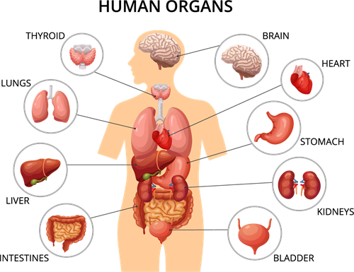
Human organs
Important!
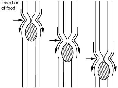
What is peristaltic movement?
Digested food is moving forward by contracting and relaxing the smooth muscles to enable nutrients to be absorbed.
Now, let's learn about one important organ, which helps us to view things.
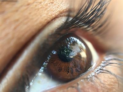
The pattern of human eye
The Eyes - Photoreceptor:
Introduction:
Our eyes belong to a group of eyes called "camera-types" (just like a camera focuses light onto a film, a structure in our eye called the cornea focuses the light onto a thin membranous structure called the retina, which helps us for a vision, differentiation of colour and to maintain our biological clock patterns.
Our eye can able to differentiate approximately 10 - 12 million colours.
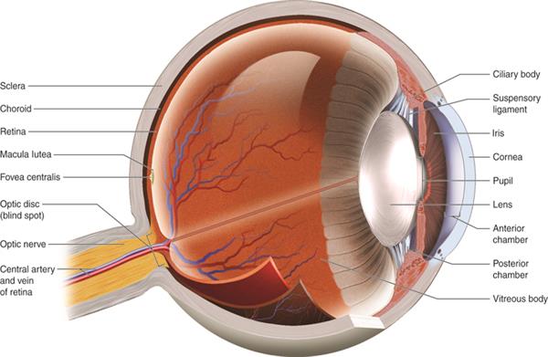
Anatomy of eye
The parts of the eye can be differentiated into two types:
1. External structure
2. Internal structure
Both structures are comprised of different types of parts and are as follows:
1. External structure:
1. External structure
2. Internal structure
Both structures are comprised of different types of parts and are as follows:
1. External structure:
Parts are as follows: sclera, conjunctiva, cornea, iris, pupil.
- Sclera: The white part of the eye protects from the outside.
- Conjunctiva:Thin outer layer protects the sclera and helps our eyes to keeps moist by secreting mucus and tears and keeps out debris enters to eyes.
- Cornea: The other transparent sheet spreads over the iris and pupil from outside and refract the light that enters the eyes.
- Iris: Iris is a ring-shaped structure located behind the cornea. The colour of human eyes are based on the pigments in the iris. The main function of the iris is to control the size of the pupil based on the light entering to eye.
- Pupil: The small opening which allows light to enter the eyes.
2. Internal structure:
Parts are as follows: lens, retina, optic nerve, aqueous humour, vitreous humour.
- Lens: It's a biconvex, adjustable crystalline part of the eye, which is made of proteins. It creates an image by collaborating with the cornea and retina.
- Retina: The light-sensitive part, which consists of the photoreceptors called rods (black & white sensitive), cones (colour sensitive), helps us to visualize the different types of images. It converts the light into electric impulses, which travels to the brain by the path of nerves called the optic nerve.
- Optic nerve: The vision would be impossible without this motor neuron (takes impulses from organs to the central nervous system - CNS) that carries the impulses of converted light by the retina to the brain (CNS).
- Aqueous Humour: The fluid which runs in between the lens and the cornea. It nourishes both parts and maintains the pressure of the eye.
- Vitreous Humour: The semi-solid, jelly-like fluid runs in between the lens and the retina, which maintains a spherical shape of the eyes and transmits light to the retina.
Reference:
https://upload.wikimedia.org/wikipedia/commons/e/ef/2404_PeristalsisN.jpg
https://courses.lumenlearning.com/austincc-ap1/chapter/special-senses-vision/
https://pixahive.com/photo/human-eye-10/
 Register for free to see more content