
PUMPA - SMART LEARNING
எங்கள் ஆசிரியர்களுடன் 1-ஆன்-1 ஆலோசனை நேரத்தைப் பெறுங்கள். டாப்பர் ஆவதற்கு நாங்கள் பயிற்சி அளிப்போம்
Book Free DemoBased on the shape of the cells and their arrangement, epithelial tissues further classified as follows:
A. Covering epithelial tissue
B. Glandular epithelial tissue
A. Covering epithelial tissue:
Covering epithelial tissue located over the surface of the body parts, both externally and internally. Based on the number of cell layers, it is classified into,
- Simple epithelial tissue
- Stratified epithelial tissue
1. Simple epithelial tissue:
It consists of a single layer of cells that forms the lining for the body cavities and ducts.
Location: It is present on the secretory and absorptive surfaces.
Based on the structure and modification of cells, simple epithelium is further classified into the following types
- Squamous epithelium
- Cuboidal epithelium
- Columnar epithelium
- Ciliated epithelium
a. Squamous epithelium:
- It is a single-layered epithelium.
- The cells are thin and flat, with disc-shaped prominent central nuclei and sparse cytoplasm with irregular boundaries.
- Cells are bind with neighbouring cells.
- It is also called pavement membrane due to its tile-like appearance.
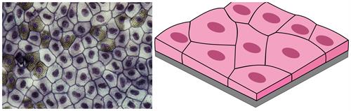
Microscopic view and illustration of the simple squamous epithelial tissue
Location: It is located in the delicate lining of the mouth, lungs' air sacs, lining of the heart, proximal tubule of kidneys, blood vessels. It also covers the skin and tongue.
Function:
- This cells protect the body from mechanical injury, drying and invasion of microbial infections.
- It helps in filtration by forming a selectively permeable membrane surface.
b. Cuboidal epithelium:
It comprises a single layer of cube-shaped cells with round nuclei, and it lies in the centre.
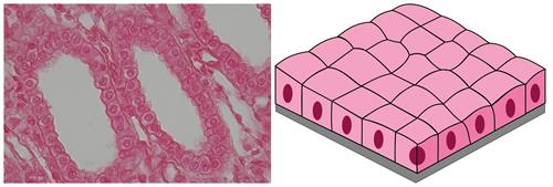
Microscopic view and illustration of the simple cuboidal epithelial tissue
Location: Cuboidal epithelium is found in the thyroid vesicles, ducts of salivary glands, sweat glands, exocrine pancreas, intestine and kidney tubules, i.e. tubular part of the nephron.
Function:
- Cuboidal epithelial cells present in the intestine and kidney tubules act as microvilli; they increase the absorption's surface area.
- The major function of the epithelial cell is the secretion of gastric juices.
Note: The functional unit of kidney is Nephron.
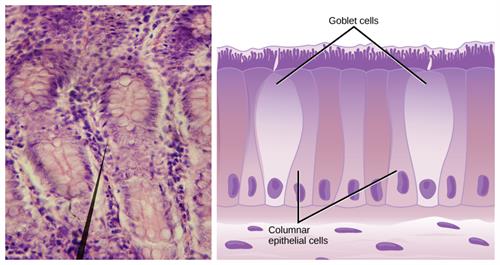
c. Columnar epithelium:
It comprises a single layer of tall, elongated, slender and pillar-like cells with oval nuclei situated at the base.
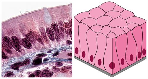
Microscopic view and illustration of the simple columnar epithelial tissue
Location: It is usually present in the stomach, and small intestine's inner lining, gall bladder, bile duct, colon, oviducts and forms the mucous membrane.
Function: These cells are mainly involved in absorbing the nutrient from the digested food and secretion of mucus and enzymes.
d. Ciliated columnar epithelium:
If the columnar epithelial cells have cilia (hair-like projections), it is called ciliated columnar epithelium.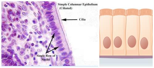
Microscopic view and illustration of the simple ciliated columnar epithelial tissue
Location: It is found in the bronchioles respiratory tract, trachea or windpipe and in the lines of fallopian tubes of oviducts and kidney tubules.
Function: Cilia can move, and their movement pushes substances like mucus forward to clear it from the ducts. The beating of the cilia moves solid dust particles in one direction through ducts.
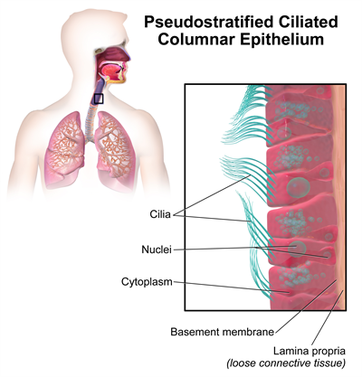
Pseudostratified ciliated columnar epithelium
B. Glandular epithelium:
Glands carry out the secretion of useful substances such as enzymes and hormones in an animal body. These are developed from epithelial tissue.
Due to the modification of epithelial cells, specialised gland cells are formed. Sometimes, a portion of epithelial cells folds inward, resulting in a multicellular gland's formation. It is called glandular epithelium.
Microscopic view and illustration of the glandular epithelium
Location: It is found in the gastric glands, intestinal glands and pancreatic tubules.
Function: It involves the synthesis and secretion of chemical substances at the epithelial surface.
Compound epithelium:
It is composed of more than one layer of cells and has limited secretion in secretion and absorption. Due to the appearance of stratified; hence it is known as stratified epithelial cells.
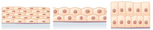
Left to right: Stratified squamous, Stratified squamous and stratified columnar
Function:
- It protects the underlying tissues against mechanical and chemical stress.
- They cover the dry surface area of the skin and moist surface area such as the buccal cavity and pharynx.
Important!
Based on the epithelial layers
- Simple - Single layer
- Stratified - More than one layer
- Ciliated - Cells with cilia
Based on the cell shapes
- Squamous - Flat
- Cuboidal - Cube
- Columnar - Rectangular
Reference:
https://upload.wikimedia.org/wikipedia/commons/e/e6/Epithelial_Tissues_Simple_Squamous_Epithelium_%28frog%29_%2827847646938%29.jpg
https://upload.wikimedia.org/wikipedia/commons/9/9e/Epithelial_Tissues_Simple_Cuboidal_Epithelium_%2841681552782%29.jpg
https://www.flickr.com/photos/146824358@N03/27854453998/
https://commons.wikimedia.org/wiki/File:Glandular_epithelium_of_small_intestine_(crypts_of_Lieberk%C3%BChn).jpg
https://commons.wikimedia.org/wiki/File:Simple_squamous_epithelium.svg
https://commons.wikimedia.org/wiki/File:Simple_cuboidal_epithelium.svg
https://commons.wikimedia.org/wiki/File:Simple_columnar_epithelium_tissue.svg
https://commons.wikimedia.org/wiki/File:Simple_columnar_epithelium.png
https://commons.wikimedia.org/wiki/File:Figure_33_02_04.png
https://commons.wikimedia.org/wiki/File:Stratified_squamous_epithelium.png
https://commons.wikimedia.org/wiki/File:Stratified_cuboidal_epithelium.png
https://en.wikipedia.org/wiki/Stratified_columnar_epithelium#/media/File:Stratified_colunar_epithelium.png
https://commons.wikimedia.org/wiki/File:Blausen_0750_PseudostratifiedCiliatedColumnar.png
 Register for free to see more content