PDF chapter test TRY NOW
3. Blood platelets:
Have you noticed that whenever an injury happens, there may be bleeding (outflow of blood) but eventually, at a certain point, the bleeding stops?
This process of stopping blood outflow is known as blood clotting and is due to blood platelets or thrombocytes.
- Platelets are tiny blood cells.
- Cells present only in mammals.
- Per cubic mm of blood contains \(2-2.5 lakhs\) of blood platelets.
- Cells are highly contractile, round, oval or biconvex with granular cytoplasm.
- The nucleus is absent.
- The life span is about \(8-10\) days.
- It is also called \(thrombocytes\).
- These cells are responsible for \(blood clotting\).

Platelet structure
Mechanism of blood clotting:
\(\huge{\downarrow}\)
Prothrombin activator converts prothrombin to thrombin.
\(\huge{\downarrow}\)
Thrombin, in turn, converts fibrinogen into fibrin.
\(\huge{\downarrow}\)
Fibrin forms a mesh-like structure that traps red blood cells.
\(\huge{\downarrow}\)
The bleeding stops when a clot forms.
Important!
Note:
- A decrease in circulating blood platelets count results in a condition called thrombocytopenia.
- Haemophilia is a condition where the thrombocytes is absent in a persons blood. Haemophiliacs (person affected by this disorder) must be extremely careful not to sustain bleeding wounds as they can bleed themselves to death.
Lymph - a white connective tissue:
- It is a colourless fluid filtered out of the blood capillaries.
- It contains plasma and white blood cells (mostly lymphocytes).
- It is a tissue that lack of red corpuscles, platelets and proteins.
- It also has lessphosphorus and calcium than the blood.
- It can clot like blood.
Function:
- Lymph mainly helps in the exchange of materials between blood and tissues.
- It also helps to destroy the invasion of pathogens.
Lymphocytes origin, maturation, and proliferation all take place in lymphoid organs.
They are classified into two types.
- Primary lymphoid organs
- Secondary lymphoid organs
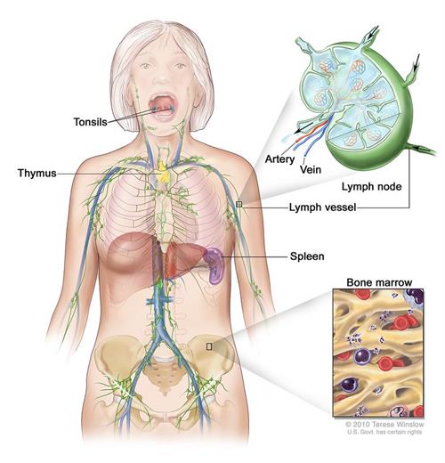
Primary and secondary lymphoid organs present in the human body
Primary lymphoid organs:
We are all familiar with lymphocytes, which are a type of white blood cells. Do you have any idea? Where did it originate?
Primary lymphoid organs are the production site of lymphocytes.
The primary lymphoid organs are,
- Bone marrow
- Thymus
In the bone topic itself, we have studied the presence of red and yellow bone marrows in long bones. Redbone marrow is the place where lymphocytes are produced. In the thymus, these lymphocytes will mature.
Secondary lymphoid organs:
The lymphocytes mature in the primary lymphoid organ, and then they transfer to a secondary lymphoid organ viz.,
- Payer's spleen
- Lymph nodes
- Tonsils
- Small intestine
- Appendix patches
These organs are the sites where the antigens will interact with lymphocytes.
Note:
An antigen is a substance that enters the body and starts a process that can cause disease. The body then usually produces substances (antibodies) to fight the antigens.
An antigen is a substance that enters the body and starts a process that can cause disease. The body then usually produces substances (antibodies) to fight the antigens.
Reference:
https://www.flickr.com/photos/nihgov/28876155584
https://upload.wikimedia.org/wikipedia/commons/c/c5/Platelet_structure.png
 Register for free to see more content