
PUMPA - SMART LEARNING
எங்கள் ஆசிரியர்களுடன் 1-ஆன்-1 ஆலோசனை நேரத்தைப் பெறுங்கள். டாப்பர் ஆவதற்கு நாங்கள் பயிற்சி அளிப்போம்
Book Free DemoJust like humans, rabbits also respire oxygen and expire carbon dioxide. These gases are exchanged into the blood at the lungs of the mammal. Since respiration processes are similar to humans, rabbits are used as animals models to validate medications for human diseases.
This object explains the respiration system of rabbits.
Respiratory canal:
Respiratory passage or canal of rabbit consists of the following parts:
- External nares
- Nasal chambers
- Internal nares
- Pharynx
- Glottis
- Larynx
Respiratory path
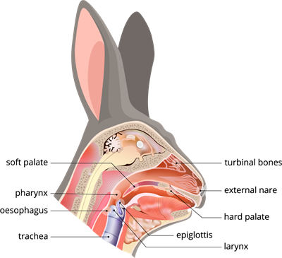
External nares:
A pair of external nares positioned at the tip of the snout take in the fresh air. The pharynx receives atmospheric air through the external nostrils and nasal tubes.
Nasal chambers:
The nasal chambers receive air from the external nares. Both nasal chambers are lined or covered by vascular mucous olfactory epithelium and have thin scroll-like turbinal bones that increase the surface area of the nasal chambers. It acts as an organ of smell.
The air becomes wet and warm as it flows through the nasal chambers. Dust particles cling to the hairs in the proximal region of the nose passage and do not reach it into the lungs.
Internal nares:
Both nasal chambers open into the nasopharynx via the internal nares, which are located above the soft palate. The nasopharynx leads to the laryngopharynx.
Pharynx:
The pharynx receives atmospheric air through the external nostrils and nasal tubes. The pharynx, a funnel-shaped passage, acts as a passageway between the respiratory and digestive tracts.
Glottis:
The glottis, a median, vertical slit-like aperture on the floor of the laryngopharynx, leads into the larynx. The epiglottis is a bilobedcartilaginous flap covered with mucous membrane that originates from the glottis' anterior margin. It covers the glottis when food is swallowed.
Air travels from the pharynx to the windpipe via the glottis.
Larynx:
The larynx, or voice box, is formed by the enlargement of the anterior part of the windpipe and its wall is supported by four cartilaginous plates. The vocal cord is located inside the larynx, and its vibrations produce sound. Rabbits don't produce loud sounds. The larynx leads into the trachea or windpipe.
Respiratory organs:
Trachea:
The trachea, or windpipe, is a long tube that runs through the neck slightly ventral to the oesophagus and continues posteriorly as the larynx. The epiglottis prevents food from entering the trachea via the glottis. The walls of the trachea are supported by cartilage rings, which aid in the free movement of air. The trachea is divided into two branches termed bronchi, with each one enters each lung and separates into bronchioles, which lead to alveoli.
Lungs:
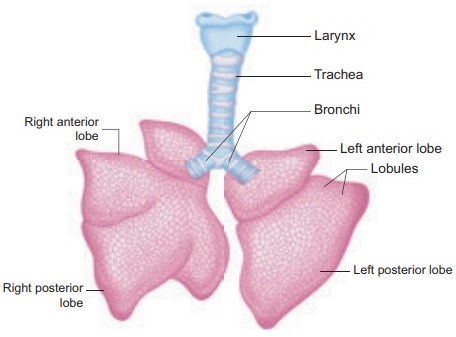
Lungs are made up of light, spongy tissues where respiration takes place. A double membrane pleura surrounds each lung. Lungs are enclosed in the thoracic cavity. Each lung is suspended from the point where the bronchus enters into it. At this point, the pulmonary artery, vein, and nerves also enter the lungs. The right lung of the rabbit is divided into four lobes, and the left lung of the rabbit has two lobes.
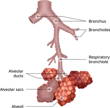
Bronchial tree
Each bronchus enters its respective lung, divides into smaller secondary and tertiary bronchi. Bronchioles are their finer terminal branches with a smaller diameter. There are no cartilage rings in the bronchioles. Each bronchiole divides again to generate finer branches called alveolar ducts, which terminate into small dilated blind air-sacs.
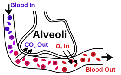
Gas exchange in alveoli
Each air sac's wall is evaginated, resulting in hollow air cells known as alveoli. Alveoli have very thin walls made of elastic connective tissue fibres and are surrounded by a network of the pulmonary artery and vein capillaries. Direct gaseous exchange occurs here because the walls of the alveoli are densely packed with blood capillaries.
Thoracic cavity of rabbit
Picture \(1\) indicates the skeletal system of the rabbit.
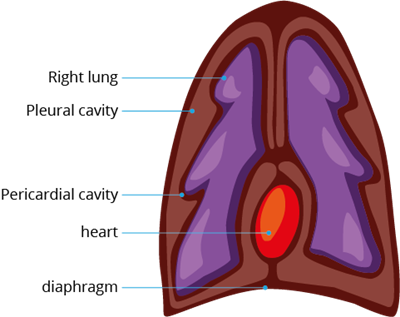
Picture \(2\) indicates the different cavities of the rabbit.
Picture \(2\) indicates the different cavities of the rabbit.
In picture \(1\), you can see the vertebral column, ribs and sternum.
- The vertebral column bound the thoracic cavity dorsally
- Sternum bound the thoracic cavity ventrally
- Ribs bound the thoracic cavity laterally
- A dome-shaped diaphragm is on the lower side of the thoracic cavity
In picture \(2\), you can see the thoracic cavity, which encloses the pair of lungs.
Inspiration and Expiration:
Inspiration (breathing in) and expiration (breathing out) are two respiratory actions that allow gases to be exchanged (oxygen and carbon dioxide). In the alveoli, gaseous exchange occurs when inspired air comes into touch with blood capillaries through the thin wall of the alveoli. Through the thin alveolar wall, oxygen from the air diffuses into the blood and carbon dioxide from the blood diffuses out into the air.
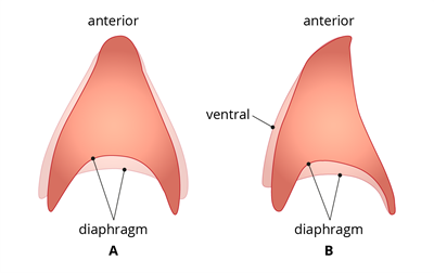
Diaphragm during breathing: A - From below: B - From side view
During inspiration, lungs will expand, which causes the air to rush into the lungs due to more outside atmospheric pressure. The outside air rushes in until the pressure of the air in the lungs equals that of the atmosphere. It is an active process.
Important!
During expiration, the lungs elastic walls contract, expelling the breathed air through the same path as before. It is a passive process.
Reference:
https://www.bioscience.com.pk/topics/zoology/item/327-breathing-and-mechanism-of-gas-exchange-respiration-in-rabbit
https://upload.wikimedia.org/wikipedia/commons/f/f9/Anatomy_and_physiology_of_animals_body_cavities.jpg
https://upload.wikimedia.org/wikipedia/commons/thumb/8/8b/Alveoli.svg/512px-Alveoli.svg.png
https://upload.wikimedia.org/wikipedia/commons/f/f9/Anatomy_and_physiology_of_animals_body_cavities.jpg
 Register for free to see more content