PDF chapter test TRY NOW
In lower organisms such as protozoans, nutrients and other materials are transported to the different parts of the cell is by means of diffusion.
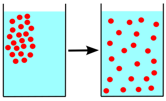
Diffusion of substances from higher to lower concentration
But in higher multicellular organisms, nutrients transfer across different parts of the organism is not possible by means of diffusion. So, a special transport system is in place for these organisms called a circulatory system. In this object circulatory system of the rabbit is explained.
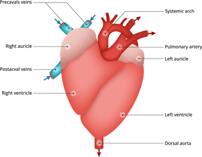
Structure of rabbit heart
Terms related to the circulatory system:
| Arteries |
|
| Veins |
|
| Pulmonary artery |
|
| Pulmonary vein |
|
| Aorta |
|
Blood, blood arteries, and the heart make up the circulatory system. The heart is pear-shaped and sits between the lungs in the thoracic cavity. Pericardium, a double-layered membrane, surrounds it.
The external and internal structure of the heart:
The heart is located in the middle space, somewhat to the left side of the thoracic cavity, between the two lungs. The heart is a muscular, four-chambered, pear-shaped pumping organ. The sharp apex of the heart points backwards, while the broad base points forward.
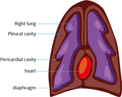
Location of heart in between the lungs
The pericardium is a thin-walled, double-layered membranous sac that entirely encloses the heart. Between the pericardium's outer parietal and inner visceral layers is a thin gap known as the pericardial cavity, which is filled with aqueous pericardial fluid.
The pericardial fluid shields the heart from damage from the outside world and allows it to move freely.
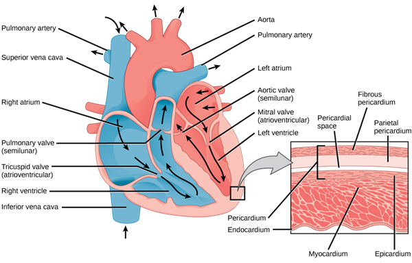
Internal structure of mammals heart with pericardium and pericardial space
The internal structure of the heart:
The internal structure of the heart consists of four chambers.
- Anterior two auricles
- Posterior two ventricles
Auricles:
The right and left auricles make up the auricular portion. The auricle on the left is smaller than the one on the right. An inter-auricular septum separates the right and left auricles. The right auricle gets deoxygenated blood from all areas of the body via two precaval (superior vena cava) and one postcaval (inferior vena cava) vein. The pulmonary veins in the lungs deliver oxygenated blood to the left auricle.
Auriculo-ventricular aperture:
Through an aperture called auriculo-ventricular aperture, both auricles open into the ventricle on their respective side. The right auriculoventricular aperture connects the right auricle to the right ventricle and is protected by the tricuspid valve. The left auriculoventricular aperture, protected by a bicuspid or mitral valve, allows the left auricle to enter the left ventricle.
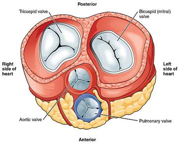
Cardiac valves of mammals
Ventricles:
Like the auricles, ventricles are divided into two chambers. An interventricular septum separates the right and left ventricles.
The pulmonary trunk emerges from the right ventricle, carrying deoxygenated blood to the lungs. The systemic arch (aorta) emerges from the left ventricle, supplying oxygenated blood to all regions of the body.
Three semilunar valves protect the openings of the pulmonary artery and aorta.
The working mechanism of the heart:
Throughout the life of the rabbit, the heart functions as a force pump, beating continually. It operates as a suction pump, sucking blood into the auricles while pumping blood into the arteries from the ventricles. The auricles contract, and the ventricles relax or dilate during heartbeats, and then the auricles relax and the ventricles contract.
Contraction and relaxation happen at the same time. Systole is the contraction, and diastole is the relaxation. The heart muscles are of the cardiac type, which has a natural tendency to beat continuously.
Through precaval and postcaval veins, the right auricle gets venous or impure blood from various areas of the body. Through pulmonary veins, oxygenated or pure blood is sent to the left auricle. The contraction of the auricles forces venous and oxygenated blood into their respective chambers of the ventricles.
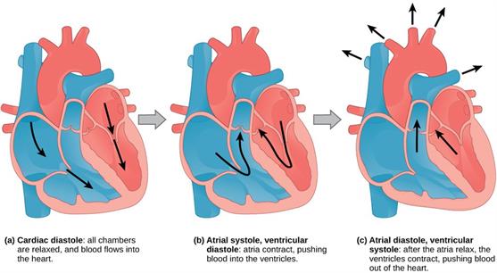
Systole and diastole of mammals heart
At the same time, both auricles contract and the right and left auriculo-ventricular apertures tricuspid and bicuspid valves are forced to open towards the ventricles, driving venous blood from the right auricle into the right ventricle and oxygenated blood from the left auricle into the left ventricle.
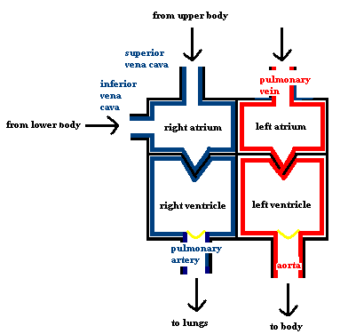
Circulation of blood in mammals
Then, a bit later than the auricles, the ventricles contract. The tricuspid and bicuspid valves close the right and left auriculo-ventricular openings when the ventricles begin to contract. As a result, blood cannot return to the auricles.
The aortic arches semilunar valves are still closed, and blood flow is impeded in all directions, increasing blood pressure inside the ventricles. When this pressure exceeds the arteries, the aortic arches' semilunar valves open, forcing blood into them.
Thus, oxygenated blood from the left ventricle travels to various areas of the body, while venous blood from the right ventricle travels to the pulmonary aorta, where it is oxygenated. The ventricles then relax, and the auricles fill with blood once more.
A heart's complete contraction (systole) and relaxation (diastole) is called a heartbeat or heart cycle.
Heartbeat
Reference:
https://upload.wikimedia.org/wikipedia/commons/f/f9/Anatomy_and_physiology_of_animals_body_cavities.jpg
https://commons.wikimedia.org/wiki/File:Diffusion.svg
https://upload.wikimedia.org/wikipedia/commons/6/62/Mammalian_Heart_and_Circulation.PNG
https://upload.wikimedia.org/wikipedia/commons/thumb/b/b9/2011_Heart_Valves.jpg/512px-2011_Heart_Valves.jpg
https://upload.wikimedia.org/wikipedia/commons/1/12/CG_Heart.gif
https://upload.wikimedia.org/wikipedia/commons/thumb/1/16/Figure_40_03_03.jpg/1024px-Figure_40_03_03.jpg
https://upload.wikimedia.org/wikipedia/commons/5/5f/Figure_40_03_02ab.png
 Register for free to see more content